
LAMENESS EXAMINATIONS
Just as in humans, orthopedic problems can cause your horse discomfort and interfere with its ability to be ridden or even to perform daily activities. Injuries to the musculoskeletal system—including bones, joints, tendons, ligaments, and muscles—can result in mild to severe lameness. Lameness is generally defined as an abnormal gait. In horses, lameness most often results from pain and much less often results from mechanical or neurologic problems.
The goal of a lameness exam is to localize the area of discomfort that results in the abnormal gait and, if possible, to identify a specific injury or disease of structures in that area that are causing this discomfort. Except in severe cases that present with obvious swelling or wounds, lameness examinations may take several steps to logically and methodically shed light on the underlying cause; our horses cannot tell us where it hurts! Regardless of the cause of lameness, early diagnosis generally gives a much better chance at successful treatment.
Any horse that cannot bear weight on a leg or that is lame at the walk should be immediately evaluated by a veterinarian. A more subtle lameness, while likely not an emergency, should be investigated promptly to ensure the best possible outcome. Please schedule ample time to be present for the veterinarian to thoroughly examine your horse. Also please understand that it may take your veterinarian more than one examination or one day of diagnostics to adequately investigate a lameness problem. If you have been giving an anti-inflammatory medication prior to the lameness exam, please ask your vet whether to stop giving it a day or two in advance to ensure the exam is accurate.
PARTS OF LAMENESS EXAM:
- Thorough discussion of horse’s medical history and of the history of the current problem. How long has it been going on? Has it improved over time? Was an incident or time of injury noted? Has the horse been on any medications?
- Examination and palpation at rest. Your vet will note any conformational abnormalities or abnormal stance. He/she may also palpate structures on the horse’s legs and body to check for swelling, any abnormalities, and reactions which may indicate discomfort.
- Hoof testers: Your vet may squeeze on the soles of each hoof with an instrument designed to check for sensitivity and pain.
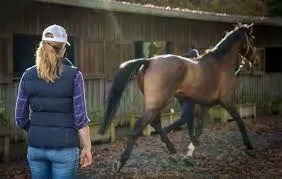
- Movement examination: Your vet will watch the horse at the walk, trot, and possibly the canter in various situations to most effectively identify the primary area of lameness. If the horse is limping, this usually means deciding on which leg the horse is limping most. The vet may watch a horse move in a straight line, on a longe line, on a hard surface, on a soft surface, performing gait transitions, and being ridden under saddle; all these circumstances may help characterize the lameness better.
- Flexion tests: Your vet will hold each leg in a flexed position for a predetermined amount of time, then watch the horse trot away and back. The response to flexion (or to standing with more weight on the opposite leg) may help identify discomfort associated with a group of joints or soft tissue structures.
DIAGNOSTIC ANALGESIA:
If a lame leg has been identified and the source of pain is not obvious, your veterinarian will likely recommend diagnostic analgesia. Analgesia means the inability to feel pain. In this step, a local anesthetic is injected around peripheral nerves or directly into synovial structures (joints and tendon sheaths) to remove sensation from the area. If the horse becomes sound after the area is desensitized, then the lameness has been localized to that area. Performing diagnostic analgesia (also called “blocking out” a lameness) requires a serious enough and consistent enough lameness (usually a grade 2 or 3) for the vet to be able to see an improvement in gait after the blocks.
In the forelimbs, vets will usually start by desensitizing most of the hoof and then moving progressively higher up the leg to systematically detect where the lameness is coming from. Many horses tolerate blocking quite well. The best analogy to a common human experience is receiving numbing injections from a dentist. If the horse does not tolerate the procedure well, then the vet may sedate your horse, perform the block, wait for the horse to become alert after sedation, and then watch the horse move again. Blocking out a lameness will direct the vet to the region of the horse of which she or he will take diagnostic images.
DIAGNOSTIC IMAGING:
Diagnostic imaging may be performed based on diagnostic analgesia (mentioned above) or based on prior findings during the lameness examination. It is important to localize the region of lameness before performing diagnostic imaging; many horses will have abnormal imaging findings that are not associated with pain (e.g. from an old injury), and localizing the lameness will increase confidence that a particular imaging finding is significant.
The imaging modalities that we have available on the farm include digital radiography (x-rays) and ultrasonography. Radiographs, or x-rays, enable us to look primarily at the bony structures in the horse’s legs, back, neck, or head. Horses are so large that radiography of the pelvis and high shoulder region is very challenging unless the horse is anesthetized. Even if radiographs are normal, your vet may recommend repeating them in a certain period of time since bony changes visible radiographically may lag behind signs of lameness with recent injuries. A digital x-ray system will display images immediately so your vet can make sure positioning is correct. Detailed evaluation of the images may be best performed back at our clinic with specialized viewing software.
Ultrasonography enables your veterinarian to examine soft-tissue structures, including tendons, ligaments, muscles, and joints. Ultrasonography can also help us look at the surfaces of bones. Your vet may compare structures on the lame leg to structures on the opposite, sound leg. Ultrasound images are best evaluated in “real-time” and are dependent on the positioning of the equipment and the technique of the sonographer.
FURTHER DIAGNOSTICS:
Depending on the course of the lameness examination and preliminary imaging, your vet may recommend further imaging with specialized modalities performed at a university hospital. These modalities include nuclear scintigraphy, computed tomography (CT), magnetic resonance imaging (MRI), and arthroscopy.
TREATMENT:
Once a cause of lameness has been identified, your vet can help you come up with a treatment plan that works for you and your horse. There are often a variety of options that may be appropriate for horses in different life stages or roles. Examples of treatments for musculoskeletal problems include:
- Joint injections with anti-inflammatories or autogenous products (e.g. interleukin 1 receptor antagonist protein (IRAP))
- Extracorporeal shockwave therapy of an injured tendon or ligament
- Injection of platelet rich plasma (PRP), another autogenous product, into a core lesion of an injured tendon or ligament
- Stall rest, small paddock turnout, and rehabilitation exercise schedules
- Topical or oral anti-inflammatory treatment
Recheck lameness examinations are important to determine degree of healing, to gauge response to treatment, and to continue planning rehabilitation.